The knee joint consists of an articulation between four bones: the femur, tibia, fibula and patella. There are four compartments to the knee. These are the medial and lateral tibio-femoral compartments, the patella-femoral compartment and the superior tibio-fibular joint. The components of each of these compartments can suffer from repetitive strain, injury or disease.
Around the knee there are primary two groups of muscle act on knee: Extensors or quadriceps muscles present at front of the thigh and flexor or hamstring muscle present at the back of the thigh.
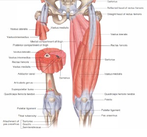
Extensors: The quadriceps muscle is the primary extensor of the knee and consists of four muscles: vastus intermedius, vastus lateralis, vastus medialis, and rectus femoris. The rectus femoris is the only one of these muscles that crosses both the hip and knee, allowing it to joint also act as a hip flexor.
Impairments– Quadriceps muscle impairments related to knee pain may include poor performance or over recruitment. The oblique portion of the vastus medialis (VMO) may provide an important restraint to lateral patellar displacement given the angulation of the fibers that attach to the patella. Poor performance of the VMO may contribute to excessive lateral patellar displacement. With intensive physical training or loading through weight training or sports, the quadriceps may become relatively too stiff or short and contribute to excessive stress on the patella, patellar ligament, or tibial tubercle. This excessive pull superiorly may be associated with patella alta. In contrast, the quadriceps can become functionally weak when habitual alignment of knee hyperextension is present.
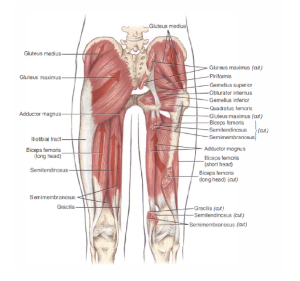
Flexors: The hamstring muscles are comprised of the semimembranosus and semitendinosus muscles medially and the biceps femoris muscle laterally. Their primary actions include knee flexion and hip extension; however, they also assist with hip medial and lateral rotation and may contribute to tibial medial and lateral rotation.
Impairments-The hamstrings are at risk of overuse when synergists, such as the gluteus maximus are underused. For example, when the foot is fixed on the ground, the hamstrings can extend the knee by pulling the knee back to the body through its action as a hip extensor. Usually, when hamstring dominance such as this occurs, the quadriceps and gluteus maximus are not performing optimally. This movement impairment is commonly seen in individuals with a swayback alignment and genu recurvatum.
The medial hamstrings assist in medial rotation of the hip and can become dominant and shorter in length compared to the biceps femoris. This imbalance in synergists is seen when an individual maintains excessive hip medial rotation or participates in sports or activities that tend to position or encourage hip medial rotation such as cycling. The biceps femoris assists in lateral rotation of the femur and lateral rotation of the tibia. When the primary hip lateral rotators are not performing optimally, the biceps femoris may compensate by increasing its action in an attempt to control hip lateral rotation. The overuse of the biceps in this way may lead to a muscle strain. The biceps femoris may also contribute to lateral rotation or posterolateral displacement of the lower leg relative to the femur, given its distal attachment to the fibular head.
The sartorius muscle flexes, abducts, and laterally rotates the hip and also flexes the knee and rotates it medially. The gracilis acts primarily to adduct the hip and also flexes and medially rotates the knee.
The gastrocnemius and soleus muscles are powerful ankle plantarflexors; however, the gastrocnemius is also a knee flexor. The plantaris also assists with ankle plantarflexion and knee flexion.
Hip muscles that can affect the knee-
The primary hip lateral rotators include the deep lateral rotators and gluteus maximus. The intrinsic or deep lateral rotators of the hip include the piriformis, gemellus superior and inferior, obturator internus and externus, and quadrates femoris muscles. As primary lateral rotators of the hip, the deep lateral rotators serve to provide precise control of rotation of the femoral head in the acetabulum, thus maintaining the integrity and stability of the hip similar to the way that the rotator cuff muscles provide control of the humeral head in the glenoid. In patients with knee pain syndromes, these muscles often become lengthened and weak, thus losing the precise control of the hip and allowing hip medial rotation to occur.
A large portion of the gluteus maximus muscle attaches into the ITB. Therefore shortness or stiffness of the gluteus maximus can contribute to relative stiffness/flexibility issues involving the ITB. Relative stiffness/flexibility as a result of shortness or stiffness of the gluteus maximus through the ITB seems to be more common in males than females and should be suspected if the patient sits in excessive hip abduction.
The secondary lateral rotators of the hip include the sartorius, the biceps femoris, and the posterior fibers of the gluteus minimus and medius. The posterior gluteus medius acts to abduct, extend, and laterally rotate the hip. The length and strength of the posterior gluteus medius is often affected by postural changes at the pelvis and hip. If an individual stands with the right iliac crest higher than the left or with the right hip in medial rotation, the posterior gluteus medius on the right may be lengthened and possibly weak, particularly if tested in the shortened position. Poor performance of this muscle is often a key factor in knee pain problems.
The TFL-ITB is a two-joint muscle complex that flexes, abducts, and medially rotates the hip and laterally rotates the tibia through its insertion onto the lateral tibial tubercle. The TFL-ITB also assists in stabilizing the knee when the knee is extended, although it does not actively extend the knee. When the TFL-ITB becomes short or too stiff, it can cause a compensatory motion such as lateral tibial rotation or valgus at the knee. The TFL-ITB can also contribute to patellar lateral glide as a result of its insertion on to the lateral patella.
The anterior fibers of the gluteus medius abduct, flex, and medially rotate the hip. Because the anterior fibers of the gluteus medius tend to be stronger than the posterior fibers, the imbalance often results in excessive medial rotation.
Causes of knee pain:
Osteoarthritis –
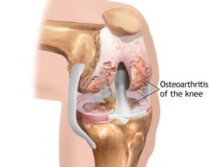
Arthritis is a complex family of musculoskeletal disorders consisting of more than 100 different diseases or conditions that destroy joints, bones, muscles, cartilage and other connective tissues, hampering or halting physical movement. Osteoarthritis (OA) is one of the oldest and most common forms of arthritis and is a chronic condition characterized by the breakdown of the joint’s cartilage. Cartilage is the part of the joint that cushions the ends of the bones and allows easy movement of joints. The breakdown of cartilage causes the bones to rub against each other, causing stiffness, pain and loss of movement in the joint.
Causes – age, obesity, muscle weakness or impairment, injury or overuse, genetic or hereditary.
Common signs and symptoms – joint soreness after periods of overuse or inactivity
Stiffness after periods of rest that goes away quickly when activity resumes
Morning stiffness, which usually lasts no more than 30 minutes
Pain caused by the weakening of muscles surrounding the joint due to inactivity
Joint pain is usually less in the morning and worse in the evening after a day’s activity
Deterioration of coordination, posture and walking due to pain and stiffness
Treatment options –
Physiotherapy treatment – To improve muscle strength and reduce pain , to improve functional capabilities, to address and correct muscle impairment and biomechanical alterations
Weight control
Medications – to reduce pain
Surgery – in severe cases
Chondromalacia patella –
Chondromalacia is due to an irritation of the undersurface of the kneecap. The undersurface of the kneecap, or patella, is covered with a layer of smooth cartilage. This cartilage normally glides effortlessly across the knee during bending of the joint. However, in some individuals, the kneecap tends to rub against one side of the knee joint, and the cartilage surface become irritated, and knee pain is the result.
Bursitis –
Prepatellar bursitis (also known as beat knee, carpet layer’s knee, coal housemaid’s knee, rug cutter’s knee, or nun’s miner’s knee, knee) is an inflammation of the prepatellar bursa at the front of the knee. It is marked by swelling at the knee, which can be tender to the touch but which does not restrict the knee’s range of motion. It is most commonly caused by trauma to the knee, either by a single acute instance or by chronic trauma over time. As such, prepatellar bursitis commonly occurs among individuals whose professions require frequent kneeling
Infrapatellar bursitis (clergyman’s knee) is the inflammation of the infrapatellar bursa, which is located just below the kneecap. It is often called “clergyman’s knee” due to its historical frequency amongst clergyman, who injured the bursa by kneeling on hard surfaces during prayer.
Tendinitis –
Patellar tendinitis (patellar tendinopathy, also known as jumper’s knee), is a relatively common cause of pain in the inferior patellar region in athletes. It is common with frequent jumping and studies have shown it may be associated with stiff ankle movement and ankle sprains.[
Some common injuries include:
- Sprain (Ligament sprain)
- Medial collateral ligament
- Lateral collateral ligament
- Anterior cruciate ligament
- Posterior cruciate ligament
- Tear of meniscus
- Medial meniscus
- Lateral meniscus
- Strain (Muscle strain)
- Quadriceps muscles
- Hamstring muscles
- Popliteal muscle
- Patellar tendon
- Hamstring tendon
- Popliteal tendon
Hemarthrosis- Hemarthrosis tends to develop over a relatively short period after injury, from several minutes to a few hours.


